Gallery - Kidney
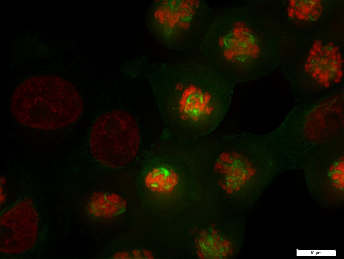
Maximum-intensity projection of a 3D widefield fluorescence image of LLC-PK1 (kidney proximal tubule) cell line from a pig kidney
- 1.42 NA oil objective lens
- 67nm pixel spacing (1376x1038 pixels)
- 13 Z slices 0.79 μm apart (under-sampled in Z)
- Staining with mCherry H2B-18 (red channel) and mEGFP Tublin-6 (green channel)
- Image dimensions (XY) are approximately 92.2 x 69.6 μm.
- Original data courtesy of Olympus Soft Imaging Solutions, Münster, Germany
Results from A Practical Guide to Deconvolution of Fluorescence Microscope Imagery, David S.C. Biggs, Microscopy Today, Volume18, Issue 01 Jan 2010, pp 10 - 14
Original
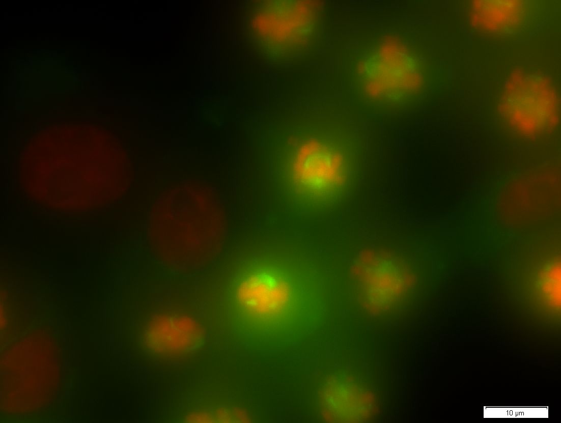
MLE (10 iterations)